


Page 175 - Textbook of Pathology, 6th Edition
P. 175
antigen of leprosy bacilli shall interact with host immune The test indicates that cell-mediated immunity is greatly 159
cells. Accordingly, the host response to the leprosy bacilli in suppressed in lepromatous leprosy while patients of
different individuals is variable. tuberculoid leprosy show good immune response. Delayed
type of hypersensitivity is conferred by T helper cells. The
3. T cell response. There is variation in T cell response in granulomas of tuberculoid leprosy have sufficient T helper
different individuals infected with leprosy bacilli: cells and fewer T suppressor cells at the periphery while the
i) Unlike tubercle bacilli, there is not only activation of CD4+ cellular infiltrates of lepromatous leprosy lack T helper cells. CHAPTER 6
T cells but also of CD8+ T cells.
ii) CD4+ T cells in lepra bacilli infected persons act not only
as helper and promoter cells but also assume the role of Classification
cytotoxicity. Leprosy is broadly classified into 2 main types:
iii) The two subpopulations of CD4+ T cells (or T helper Lepromatous type representing low resistance; and
cells)—T 1 cells and T 2 cells, elaborate different types of Tuberculoid type representing high resistance.
H
H
cytokines in response to stimuli from the lepra bacilli and
macrophages. Salient differences between the two main forms of lep-
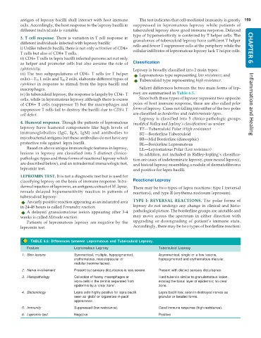
iv) In tuberculoid leprosy, the response is largely by CD4+ T rosy are summarised in Table 6.5.
cells, while in lepromatous leprosy although there is excess Since both these types of leprosy represent two opposite
of CD8+ T cells (suppressor T) but the macrophages and poles of host immune response, these are also called polar Inflammation and Healing
suppressor T cells fail to destroy the bacilli due to CD8+ T forms of leprosy. Cases not falling into either of the two poles
cell defect. are classified as borderline and indeterminate types.
Leprosy is classified into 5 clinico-pathologic groups
4. Humoral response. Though the patients of lepromatous (modified Ridley and Jopling’s classification) as under:
leprosy have humoral components like high levels of TT—Tuberculoid Polar (High resistance)
immunoglobulins (IgG, IgA, IgM) and antibodies to BT—Borderline Tuberculoid
mycobacterial antigens but these antibodies do not have any BB—Mid Borderline (dimorphic)
protective role against lepra bacilli. BL—Borderline Lepromatous
Based on above unique immunologic features in leprosy, LL—Lepromatous Polar (Low resistance)
lesions in leprosy are classified into 5 distinct clinico- In addition, not included in Ridley-Jopling’s classifica-
pathologic types and three forms of reactional leprosy which tion are cases of indeterminate leprosy, pure neural leprosy,
are described below), and an intradermal immunologic test, and histoid leprosy resembling a nodule of dermatofibroma
lepromin test. and positive for lepra bacilli.
LEPROMIN TEST. It is not a diagnostic test but is used for
classifying leprosy on the basis of immune response. Intra- Reactional Leprosy
dermal injection of lepromin, an antigenic extract of M. leprae, There may be two types of lepra reactions: type I (reversal
reveals delayed hypersensitivity reaction in patients of reactions), and type II (erythema nodosum leprosum).
tuberculoid leprosy:
An early positive reaction appearing as an indurated area TYPE I: REVERSAL REACTIONS. The polar forms of
in 24-48 hours is called Fernandez reaction. leprosy do not undergo any change in clinical and histo-
A delayed granulomatous lesion appearing after 3-4 pathological picture. The borderline groups are unstable and
weeks is called Mitsuda reaction. may move across the spectrum in either direction with
Patients of lepromatous leprosy are negative by the upgrading or downgrading of patient’s immune state.
lepromin test. Accordingly, there may be two types of borderline reaction:
TABLE 6.5: Differences between Lepromatous and Tuberculoid Leprosy.
Feature Lepromatous Leprosy Tuberculoid Leprosy
1. Skin lesions Symmetrical, multiple, hypopigmented, Asymmetrical, single or a few lesions,
erythematous, maculopapular or hypopigmented and erythematous macular.
nodular (leonine facies).
2. Nerve involvement Present but sensory disturbance is less severe. Present with distinct sensory disturbance.
3. Histopathology Collection of foamy macrophages or Hard tubercle similar to granulomatous lesion,
lepra cells in the dermis separated from eroding the basal layer of epidermis; no clear
epidermis by a ‘clear zone’. zone.
4. Bacteriology Lepra cells highly positive for lepra bacilli Lepra bacilli few, seen in destroyed nerves as
seen as ‘globi’ or ‘cigarettes-in-pack’ granular or beaded forms.
appearance.
5. Immunity Suppressed (low resistance). Good immune response (high resistance).
6. Lepromin test Negative Positive

