


Page 495 - Textbook of Pathology, 6th Edition
P. 495
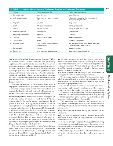
TABLE 17.5: Contrasting Salient Features of ‘Predominant Bronchitis’ and ‘Predominant Emphysema’. 479
Feature Predominant Bronchitis Predominant Emphysema
1. Age at diagnosis About 50 years About 60 years
2. Underlying pathology Hypertrophy of mucus-producing Inflammatory narrowing of bronchioles and
cells destruction of septal walls
3. Dyspnoea Late, mild Early, severe
4. Cough Before dyspnoea starts After dyspnoea starts
5. Sputum Copious, purulent Scanty, mucoid, less frequent
6. Bronchial infections More frequent Less frequent
7. Respiratory insufficiency Repeated Terminal
8. Cyanosis Common (‘blue-bloaters’) Rare (‘pink-puffers’)
9. Lung capacity Normal Increased (barrel-chest)
10. Blood gas values ↓ pO 2 , ↓ pCO 2 , no compensatory pO 2 and pCO 2 usually within normal limits due
hyperventilation to compensatory hyperventilation
11. Cor pulmonale Frequent Rare and terminal
12. Chest X-ray Large heart, prominent vessels Small heart, hyperinflated lungs
ETIOPATHOGENESIS. The commonest form of COPD is The most common abnormal phenotype in classic α1-AT
the combination of chronic bronchitis and pulmonary deficiency is homozygous state PiZZ resulting from a single
emphysema. Chronic bronchitis, however, does not always amino acid substitution Glu→Lys which causes spontaneous CHAPTER 17
lead to emphysema nor all cases of emphysema have changes polymerisation of α1-AT and inhibits its release from the
of chronic bronchitis. The association of the two conditions liver. The remaining material of α1-AT in the liver causes
is principally linked to the common etiologic factors— most hepatic cirrhosis (Chapter 22).
importantly tobacco smoke and air pollutants. Other less Clinically significant deficiency is also associated with
significant contributory factors are occupational exposure, homozygous Pi null null and heterozygous Pi nullZ.
infection and somewhat poorly-understood familial and The heterozygote pattern of PiMZ has intermediate levels
genetic influences. All these factors have already been which is not sufficient to produce clinical deficiency, but
discussed above. heterozygote individuals who smoke heavily have higher
However, pathogenesis of the most significant event in risk of developing emphysema.
emphysema, the destruction of the alveolar walls, is not linked The α1-AT deficiency develops in adults and causes The Respiratory System
to bronchial changes but is closely related to deficiency of pulmonary emphysema in smokers as well as in non-
serum alpha-1-antitrypsin (α1-protease inhibitor) commonly smokers, though the smokers become symptomatic about
termed protease-antiprotease hypothesis detailed below. 15 years earlier than non-smokers. The other organ showing
effects of α1-AT deficiency is the liver which may develop
Protease-antiprotease hypothesis. Alpha-1-antitrypsin (α- obstructive jaundice early in infancy, and cirrhosis and
1-AT), also called α1-protease inhibitor (α-1-Pi), is a hepatoma late in adulthood (Chapter 22).
glycoprotein that forms the normal constituent of the α1- The mechanism of alveolar wall destruction in
globulin fraction of the plasma proteins on serum emphysema by elastolytic action is based on the imbalance
electrophoresis. The single gene locus that codes for α-1-AT between proteases (chiefly elastase) and anti-proteases (chiefly
is located on the long arm of chromosome 15. It is normally anti-elastase):
synthesised in the liver and is distributed in the circulating By decreased anti-elastase activity i.e. deficiency of α-1
blood, tissue fluids and macrophages. The normal function antitrypsin.
of α1-AT is to inhibit proteases and hence its name α1- By increased activity of elastase i.e. increased neutrophilic
protease inhibitor. The proteases (mainly elastases) are infiltration in the lungs causing excessive elaboration of
derived from neutrophils. Neutrophil elastase has the neutrophil elastase.
capability of digesting lung parenchyma but is inhibited from There are enough evidences to suggest that smoking
doing so by anti-elastase effect of α1-AT. promotes emphysema by both decreasing the amount of anti-
There are several known alleles of α1-AT which have an elastase as well as by increasing the elastolytic protease in
autosomal codominant inheritance pattern and are classified the lungs. These are as under:
as normal (PiMM), deficient (PiZZ), null type (Pi null null) 1. Oxidant in cigarette smoke has inhibitory influence on
having no detectable level, and dysfunctional (PiSS) type α-1-antitrypsin, thus lowering the level of anti-elastase
having about half the normal level. activity.

