


Page 496 - Textbook of Pathology, 6th Edition
P. 496
480                                                        Grossly, the lungs are voluminous, pale with little blood.
                                                                 The edges of the lungs are rounded. Mild cases show
                                                                 dilatation of air spaces visible with hand lens. Advanced
                                                                 cases show subpleural bullae and blebs bulging outwards
                                                                 from the surface of the lungs with rib markings between
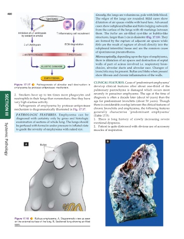
                                                                 them. The  bullae are air-filled cyst-like or bubble-like
                                                                 structures, larger than 1 cm in diameter (Fig. 17.18). They
                                                                 are formed by the rupture of adjacent air spaces while
                                                                 blebs are the result of rupture of alveoli directly into the
                                                                 subpleural interstitial tissue and are the common cause
                                                                 of spontaneous pneumothorax.
                                                                 Microscopically, depending upon the type of emphysema,
                                                                 there is dilatation of air spaces and destruction of septal
                                                                 walls of part of acinus involved i.e. respiratory bron-
                                                                 chioles, alveolar ducts and alveolar sacs. Changes of
                                                                 bronchitis may be present. Bullae and blebs when present
                                                                 show fibrosis and chronic inflammation of the walls.

                                                               CLINICAL FEATURES. Cases of ‘predominant emphysema’
           Figure 17.17  Pathogenesis of alveolar wall destruction in  develop clinical features after about one-third of the
           emphysema by protease-antiprotease mechanism.
                                                               pulmonary parenchyma is damaged which occurs most
           2. Smokers have up to ten times more phagocytes and  severely in panacinar emphysema. The age at the time of
           neutrophils in their lungs than nonsmokers; thus they have  diagnosis is often a decade later (about 60 years) than the
           very high elastase activity.                        age for predominant bronchitis (about 50 years). Though
              Pathogenesis of emphysema by protease-antiprotease  there is considerable overlap between the clinical features of
           mechanism is diagrammatically illustrated in Fig. 17.17.  chronic bronchitis and emphysema, the following features
                                                               generally characterise ‘predominant emphysema’
            PATHOLOGIC FEATURES. Emphysema can be              (Table 17.5):
     SECTION III
            diagnosed with certainty only by gross and histologic  1. There is long history of slowly increasing severe
            examination of sections of whole lung. The lungs should  exertional dyspnoea.
            be perfused with formalin under pressure in inflated state  2. Patient is quite distressed with obvious use of accessory
            to grade the severity of emphysema with naked eye.  muscles of respiration.


     Systemic Pathology


           Figure 17.18  Bullous emphysema. A, Diagrammatic view as seen
           on the external surface of the lung. B, Sectioned lung showing air-filled
           sacs.

