


Page 497 - Textbook of Pathology, 6th Edition
P. 497
3. Chest is barrel-shaped and hyperresonant.          appearance is obvious in cut surface of the lung. It shows  481
           4. Cough occurs late after dyspnoea starts and is associated  distended air spaces in the centre of the lobules
           with scanty mucoid sputum.                            surrounded by a rim of normal lung parenchyma in the
           5. Recurrent respiratory infections are not frequent.  same lobule. The lobules are separated from each other
           6. Patients are called ‘pink puffers’ as they remain well  by fine fibrous tissue septa. Large amount of black
           oxygenated and have tachypnoea.                       pigment is often present in the walls of emphysematous
           7. Weight loss is common.                             spaces. In more severe cases, distal parts of acini are also
           8. Features of right heart failure (cor pulmonale) and  involved and the appearance may closely resemble
           hypercapneic respiratory failure are the usual terminal  panacinar emphysema.
           events.
           9. Chest X-ray shows small heart with hyperinflated lungs.  Microscopically, there is distension and destruction of the
              After these general comments about morphologic and  respiratory bronchiole in the centre of lobules, surroun-
           clinical features of emphysema, the specific pathologic  ded peripherally by normal uninvolved alveoli. The
                                                                 terminal bronchioles supplying the acini show chronic
           changes in individual types of ‘emphysema’ and
           ‘overinflation’ as classified in Table 17.4 are described below.  inflammation and are narrowed.
                                                               2. PANACINAR (PANLOBULAR) EMPHYSEMA.
           Morphology of Individual Types of Emphysema         Panacinar or panlobular emphysema is the other common
                                                               type. In this type, all portions of the acinus are affected but
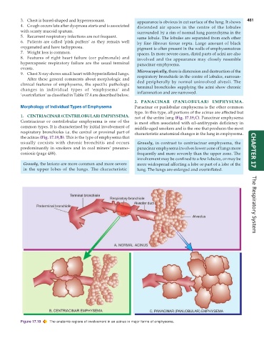
           1. CENTRIACINAR (CENTRILOBULAR) EMPHYSEMA.          not of the entire lung (Fig. 17.19,C). Panacinar emphysema
           Centriacinar or centrilobular emphysema is one of the  is most often associated with α1-antitrypsin deficiency in
           common types. It is characterised by initial involvement of  middle-aged smokers and is the one that produces the most
           respiratory bronchioles i.e. the central or proximal part of  characteristic anatomical changes in the lung in emphysema.
           the acinus (Fig. 17.19,B). This is the type of emphysema that
           usually coexists with chronic bronchitis and occurs   Grossly, in contrast to centriacinar emphysema, the
           predominantly in smokers and in coal miners’ pneumo-  panacinar emphysema involves lower zone of lungs more
           coniosis (page 488).                                  frequently and more severely than the upper zone. The  CHAPTER 17
                                                                 involvement may be confined to a few lobules, or may be
            Grossly, the lesions are more common and more severe  more widespread affecting a lobe or part of a lobe of the
            in the upper lobes of the lungs. The characteristic  lung. The lungs are enlarged and overinflated.       The Respiratory System


           Figure 17.19  The anatomic regions of involvement in an acinus in major forms of emphysema.

