


Page 498 - Textbook of Pathology, 6th Edition
P. 498
482


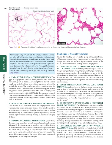
           Figure 17.20  Panacinar (Panlobular) emphysema showing involvement of the entire lobules and whole of acinus.


            Microscopically, usually all the alveoli within a lobule  Morphology of Types of Overinflation
            are affected to the same degree. All portions of acini are  Under this heading are covered a group of lung conditions
            distended—respiratory bronchioles, alveolar ducts and  of heterogeneous etiology characterised by overinflation of
            alveoli, are all dilated and their walls stretched and thin.  the parts of acini but without significant destruction of the
            Ruptured alveolar walls and spurs of broken septa are  walls and are sometimes loosely termed emphysema.
            seen between the adjacent alveoli. The capillaries are  1. COMPENSATORY OVERINFLATION (COMPEN-
            stretched and thinned. Special stains show loss of elastic  SATORY EMPHYSEMA). When part of a lung or a lobe of
            tissue. Inflammatory changes are usually absent    lung is surgically removed, the residual lung parenchyma
     SECTION III
            (Fig. 17.20).                                      undergoes compensatory hyperinflation so as to fill the
                                                               pleural cavity. Histologic examination shows dilatation of
           3. PARASEPTAL (DISTAL ACINAR) EMPHYSEMA. This       alveoli but no destruction of septal walls and hence the term
           type of emphysema involves distal part of acinus while the  compensatory overinflation is preferable over ‘compensatory
           proximal part is normal. Paraseptal or distal acinar  emphysema’.
           emphysema is localised along the pleura and along the  2. SENILE HYPERINFLATION (AGING LUNG, SENILE
           perilobular septa. The involvement is seen adjacent to the  EMPHYSEMA). In old people, the lungs become voluminous
           areas of fibrosis and atelectasis and involves upper part of  due to loss of elastic tissue, thinning and atrophy of the
           lungs more severely than the lower. This form of emphysema  alveolar ducts and alveoli. The alveoli are thin-walled and
           is seldom associated with COPD but is the common cause of  distended throughout the lungs but there is no significant
           spontaneous pneumothorax in young adults. Grossly, the  destruction of the septal walls and, therefore, preferable
     Systemic Pathology
           subpleural portion of the lung shows air-filled cysts, 0.5 to  designation is ‘senile hyperinflation’ over ‘senile
           2 cm in diameter.                                   emphysema.’

           4. IRREGULAR (PARA-CICATRICIAL) EMPHYSEMA.          3. OBSTRUCTIVE OVERINFLATION (INFANTILE
           This is the most common form of emphysema, seen     LOBAR EMPHYSEMA). Partial obstruction to the bronchial
           surrounding scars from any cause. The involvement is  tree such as by a tumour or a foreign body causes
           irregular as regards the portion of acinus involved as well as  overinflation of the region supplied by obstructed bronchus.
           within the lung as a whole. During life, irregular emphysema  Infantile lobar emphysema is a variant of obstructive
           is often asymptomatic and may be only an incidental autopsy  overinflation occurring in infants in the first few days of life
           finding.                                            who develop respiratory distress or who have congenital
                                                               hypoplasia of bronchial cartilage. In all such cases, air enters
           5. MIXED (UNCLASSIFIED) EMPHYSEMA. Quite often,     the lungs during inspiration but cannot leave on expiration
           the same lung may show more than one type of emphysema.  resulting in ballooning up of the affected part of the lung.
           It is usually due to more severe involvement resulting in loss  4. UNILATERAL TRANSLUCENT LUNG (UNILATE-
           of clearcut distinction between one type of emphysema and  RAL EMPHYSEMA). This is a form of overinflation in which
           the other. Thus, the lungs of an elderly smoker at autopsy  one lung or one of its lobes or segments of a lobe are
           may show continuation of centriacinar emphysema in the  radiolucent. The condition occurs in adults and there is
           upper lobes, panacinar in the lower lobes, and paraseptal  generally a history of serious pulmonary infection in
           emphysema in the subpleural region.                 childhood, probably bronchiolitis obliterans. The affected

