


Page 64 - Textbook of Pathology, 6th Edition
P. 64
48 6. Appearance of phosphatidylserine and thrombospondin
3. The nuclear chromatin is condensed or fragmented
(pyknosis or karyorrehexis). on the outer surface of apoptotic cell facilitates early
recognition by macrophages for phagocytosis prior to
4. The cell membrane may show convolutions or projections appearance of inflammatory cells.
on the surface.
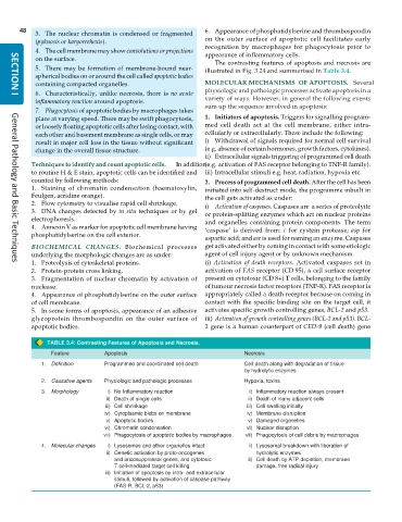
The contrasting features of apoptosis and necrosis are
5. There may be formation of membrane-bound near- illustrated in Fig. 3.24 and summarised in Table 3.4.
spherical bodies on or around the cell called apoptotic bodies
containing compacted organelles. MOLECULAR MECHANISMS OF APOPTOSIS. Several
physiologic and pathologic processes activate apoptosis in a
6. Characteristically, unlike necrosis, there is no acute variety of ways. However, in general the following events
SECTION I
inflammatory reaction around apoptosis. sum up the sequence involved in apoptosis:
7. Phagocytosis of apoptotic bodies by macrophages takes
place at varying speed. There may be swift phagocytosis, 1. Initiators of apoptosis. Triggers for signalling program-
or loosely floating apoptotic cells after losing contact, with med cell death act at the cell membrane, either intra-
each other and basement membrane as single cells, or may cellularly or extracellularly. These include the following:
result in major cell loss in the tissue without significant i) Withdrawal of signals required for normal cell survival
change in the overall tissue structure. (e.g. absence of certain hormones, growth factors, cytokines).
ii) Extracellular signals triggering of programmed cell death
Techniques to identify and count apoptotic cells. In addition (e.g. activation of FAS receptor belonging to TNF-R family).
to routine H & E stain, apoptotic cells can be identified and iii) Intracellular stimuli e.g. heat, radiation, hypoxia etc.
counted by following methods: 2. Process of programmed cell death. After the cell has been
1. Staining of chromatin condensation (haematoxylin, initiated into self-destruct mode, the programme inbuilt in
Feulgen, acridine orange). the cell gets activated as under:
2. Flow cytometry to visualise rapid cell shrinkage. i) Activation of caspases. Caspases are a series of proteolyitc
3. DNA changes detected by in situ techniques or by gel or protein-splitting enzymes which act on nuclear proteins
electrophoresis. and organelles containing protein components. The term
4. Annexin V as marker for apoptotic cell membrane having ‘caspase’ is derived from: c for cystein protease; asp for
phosphatidylserine on the cell exterior.
aspartic acid; and ase is used for naming an enzyme. Caspases
BIOCHEMICAL CHANGES. Biochemical processes get activated either by coming in contact with some etiologic
underlying the morphologic changes are as under: agent of cell injury agent or by unknown mechanism.
1. Proteolysis of cytoskeletal proteins. ii) Activation of death receptors. Activated caspases set in
General Pathology and Basic Techniques
2. Protein-protein cross linking. activation of FAS receptor (CD 95), a cell surface receptor
3. Fragmentation of nuclear chromatin by activation of present on cytotoxic (CD 8+) T cells, belonging to the family
nuclease. of tumour necrosis factor receptors (TNF-R). FAS receptor is
4. Appearance of phosphatidylserine on the outer surface appropriately called a death receptor because on coming in
of cell membrane. contact with the specific binding site on the target cell, it
5. In some forms of apoptosis, appearance of an adhesive activates specific growth controlling genes, BCL-2 and p53.
glycoprotein thrombospondin on the outer surface of iii) Activation of growth controlling genes (BCL-2 and p53). BCL-
apoptotic bodies. 2 gene is a human counterpart of CED-9 (cell death) gene
TABLE 3.4: Contrasting Features of Apoptosis and Necrosis.
Feature Apoptosis Necrosis
1. Definition Programmed and coordinated cell death Cell death along with degradation of tissue
by hydrolytic enzymes
2. Causative agents Physiologic and pathologic processes Hypoxia, toxins
3. Morphology i) No Inflammatory reaction i) Inflammatory reaction always present
ii) Death of single cells ii) Death of many adjacent cells
iii) Cell shrinkage iii) Cell swelling initially
iv) Cytoplasmic blebs on membrane iv) Membrane disruption
v) Apoptotic bodies v) Damaged organelles
vi) Chromatin condensation vi) Nuclear disruption
vii) Phagocytosis of apoptotic bodies by macrophages vii) Phagocytosis of cell debris by macrophages
4. Molecular changes i) Lysosomes and other organelles intact i) Lysosomal breakdown with liberation of
ii) Genetic activation by proto-oncogenes hydrolytic enzymes
and oncosuppressor genes, and cytotoxic ii) Cell death by ATP depletion, membrane
T cell-mediated target cell killing damage, free radical injury
iii) Initiation of apoptosis by intra- and extracellular
stimuli, followed by activation of caspase pathway
(FAS-R, BCL-2, p53)

