


Page 65 - Textbook of Pathology, 6th Edition
P. 65
found in programmed cell death of nematode worm     virulent bacteria resulting in massive tissue necrosis. Thus,  49
           Caenorabditis elegans. BCL-2 gene family is located in the outer  the end-result of necrotising inflammation and gangrene is
           mitochondrial membrane and includes both activators and  the same but the way the two are produced, is different. The
           inhibitors of apoptosis. Thus, it may regulate the apoptotic  examples of necrotising inflammation are: gangrenous
           process by binding to some related proteins (e.g. to BAX and  appendicitis, gangrenous stomatitis (noma, cancrum oris).
           BAD) for promoting apoptosis, or to BCL-XL for inhibiting  There are 2 main forms of gangrene—dry and wet, and a
           apoptosis. The net effect on the mitochondrial membrane is  variant form of wet gangrene called gas gangrene. In all types  CHAPTER 3
           thus based on the pro-apoptotic and anti-apoptotic actions  of gangrene, necrosis undergoes liquefaction by the action
           of BCL-2 gene family.                               of putrefactive bacteria.
              Besides  BCL-2,  the apoptotic pathway is partly also
           governed by p53 molecule which promotes apoptosis.  Dry Gangrene
           iv) Cell death.  The above mechanisms lead to proteolytic  This form of gangrene begins in the distal part of a limb due
           actions on nucleus, chromatin clumping, cytoskeletal  to ischaemia. The typical example is the dry gangrene in the
           damage, disruption of endoplasmic reticulum, mitochondrial  toes and feet of an old patient due to arteriosclerosis. Other
           damage, and disturbed cell membrane.                causes of dry gangrene foot include thromboangiitis
                                                               obliterans (Buerger’s disease), Raynaud’s disease, trauma,
           3. Phagocytosis. The dead apoptotic cells develop   ergot poisoning. It is usually initiated in one of the toes which
           membrane changes which promote their phagocytosis.  is farthest from the blood supply, containing so little blood
           Phosphatidylserine and thrombospondin molecules which  that even the invading bacteria find it hard to grow in the
           are normally present on the inside of the cell membrane,  necrosed tissue. The gangrene spreads slowly upwards until  Cell Injury and Cellular Adaptations
           appear on the outer surface of the cells in apoptosis, which  it reaches a point where the blood supply is adequate to keep
           facilitate their identification by adjacent phagocytes and  the tissue viable. A line of separation is formed at this point
           promotes phagocytosis. The phagocytosis is unaccompanied  between the gangrenous part and the viable part.
           by any other inflammatory cells.
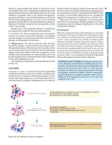
              The mechanism of apoptosis is schematically represented  MORPHOLOGIC FEATURES. Grossly, the affected part
           in Fig. 3.31.                                         is dry, shrunken and dark black, resembling the foot of a
                                                                 mummy. It is black due to liberation of haemoglobin from
           GANGRENE                                              haemolysed red blood cells which is acted upon by
                                                                 hydrogen disulfide (H S) produced by bacteria resulting
                                                                                    2
           Gangrene is a form of necrosis of tissue with superadded  in formation of black iron sulfide. The line of separation
           putrefaction. The type of necrosis is usually coagulative due  usually brings about complete separation with eventual
           to ischaemia (e.g. in gangrene of the bowel, gangrene of limb).  falling off of the gangrenous tissue if it is not removed
           On the other hand, gangrenous or necrotising inflammation is  surgically (Fig. 3.32).
           characterised by primarily inflammation provoked by


           Figure 3.31  Molecular mechanism of apoptosis.

