
Page 700 - Textbook of Pathology, 6th Edition
P. 700
684
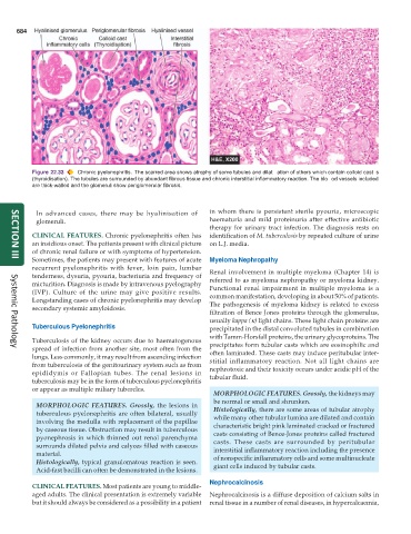
Figure 22.33 Chronic pyelonephritis. The scarred area shows atrophy of some tubules and dilat ation of others which contain colloid cast s
(thyroidisation). The tubules are surrounded by abundant fibrous tissue and chronic interstitial inflammatory reaction. The blo od vessels included
are thick-walled and the glomeruli show periglomerular fibrosis.
In advanced cases, there may be hyalinisation of in whom there is persistent sterile pyouria, microscopic
glomeruli. haematuria and mild proteinuria after effective antibiotic
therapy for urinary tract infection. The diagnosis rests on
CLINICAL FEATURES. Chronic pyelonephritis often has identification of M. tuberculosis by repeated culture of urine
an insidious onset. The patients present with clinical picture on L.J. media.
of chronic renal failure or with symptoms of hypertension.
Sometimes, the patients may present with features of acute Myeloma Nephropathy
SECTION III
recurrent pyelonephritis with fever, loin pain, lumbar
tenderness, dysuria, pyouria, bacteriuria and frequency of Renal involvement in multiple myeloma (Chapter 14) is
micturition. Diagnosis is made by intravenous pyelography referred to as myeloma nephropathy or myeloma kidney.
(IVP). Culture of the urine may give positive results. Functional renal impairment in multiple myeloma is a
Longstanding cases of chronic pyelonephritis may develop common manifestation, developing in about 50% of patients.
secondary systemic amyloidosis. The pathogenesis of myeloma kidney is related to excess
filtration of Bence Jones proteins through the glomerulus,
usually kappa (κ) light chains. These light chain proteins are
Tuberculous Pyelonephritis precipitated in the distal convoluted tubules in combination
with Tamm-Horsfall proteins, the urinary glycoproteins. The
Tuberculosis of the kidney occurs due to haematogenous
spread of infection from another site, most often from the precipitates form tubular casts which are eosinophilic and
Systemic Pathology
lungs. Less commonly, it may result from ascending infection often laminated. These casts may induce peritubular inter-
from tuberculosis of the genitourinary system such as from stitial inflammatory reaction. Not all light chains are
epididymis or Fallopian tubes. The renal lesions in nephrotoxic and their toxicity occurs under acidic pH of the
tuberculosis may be in the form of tuberculous pyelonephritis tubular fluid.
or appear as multiple miliary tubercles.
MORPHOLOGIC FEATURES. Grossly, the kidneys may
be normal or small and shrunken.
MORPHOLOGIC FEATURES. Grossly, the lesions in
tuberculous pyelonephritis are often bilateral, usually Histologically, there are some areas of tubular atrophy
involving the medulla with replacement of the papillae while many other tubular lumina are dilated and contain
by caseous tissue. Obstruction may result in tuberculous characteristic bright pink laminated cracked or fractured
pyonephrosis in which thinned out renal parenchyma casts consisting of Bence-Jones proteins called fractured
surrounds dilated pelvis and calyces filled with caseous casts. These casts are surrounded by peritubular
material. interstitial inflammatory reaction including the presence
Histologically, typical granulomatous reaction is seen. of nonspecific inflammatory cells and some multinucleate
Acid-fast bacilli can often be demonstrated in the lesions. giant cells induced by tubular casts.
Nephrocalcinosis
CLINICAL FEATURES. Most patients are young to middle-
aged adults. The clinical presentation is extremely variable Nephrocalcinosis is a diffuse deposition of calcium salts in
but it should always be considered as a possibility in a patient renal tissue in a number of renal diseases, in hypercalcaemia,