
Page 831 - Textbook of Pathology, 6th Edition
P. 831
815
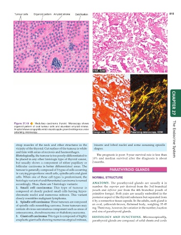
Figure 27.19 Medullary carcinoma thyroid. Microscopy shows CHAPTER 27
organoid pattern of oval tumour cells and abundant amyloid stroma.
Amyloid shows congophilia which depicts apple-green birefringence under
polarising microscopy.
strap muscles of the neck and other structures in the bizarre and lobed nuclei and some assuming spindle The Endocrine System
vicinity of the thyroid. Cut surface of the tumour is white shapes.
and firm with areas of necrosis and haemorrhages.
Histologically, the tumour is too poorly-differentiated to The prognosis is poor: 5-year survival rate is less than
be placed in any other histologic type of thyroid cancer, 10% and median survival after the diagnosis is about
but usually shows a component of either papillary or 2 months.
follicular carcinoma in better differentiated areas. The
tumour is generally composed of 3 types of cells occurring PARATHYROID GLANDS
in varying proportions: small cells, spindle cells and giant
cells. When one of these cell types is predominant, the NORMAL STRUCTURE
histologic variant of undifferentiated carcinoma is named
accordingly. Thus, there are 3 histologic variants: ANATOMY. The parathyroid glands are usually 4 in
1. Small cell carcinoma: This type of tumour is number: the superior pair derived from the 3rd branchial
composed of closely packed small cells having hyper- pouch and inferior pair from the 4th branchial pouch of
chromatic nuclei and numerous mitoses. This variant primitive foregut. Both pairs are usually embedded in the
closely resembles malignant lymphoma. posterior aspect of the thyroid substance but separated from
2. Spindle cell carcinoma: These tumours are composed it by a connective tissue capsule. In the adults, each gland is
of spindle cells resembling sarcoma. Some tumours may an oval, yellowish-brown, flattened body, weighing 35-45
contain obvious sarcomatous component such as areas of mg. There may, however, be variation in the number, location
osteosarcoma, chondrosarcoma or rhabdomyosarcoma. and size of parathyroid glands.
3. Giant cell carcinoma: This type is composed of highly HISTOLOGY AND FUNCTIONS. Microscopically,
anaplastic giant cells showing numerous atypical mitoses, parathyroid glands are composed of solid sheets and cords