
Page 838 - Textbook of Pathology, 6th Edition
P. 838
822 identified. Currently, it is proposed that insulin resistance 3. Two main mechanisms for hyperglycaemia in type 2 DM—
may be possibly due to one of the following defects: insulin resistance and impaired insulin secretion, are interlinked.
Polymorphism in various post-receptor intracellular signal 4. While obesity plays a role in pathogenesis of insulin
pathway molecules. resistance, impaired insulin secretion may be from many
Elevated free fatty acids seen in obesity may contribute e.g. constitutional factors.
by impaired glucose utilisation in the skeletal muscle, by 5. Increased hepatic synthesis of glucose in initial period of
increased hepatic synthesis of glucose, and by impaired disease contributes to hyperglycaemia.
β-cell function.
Insulin resistance syndrome is a complex of clinical features Morphologic Features in Pancreatic Islets
occurring from insulin resistance and its resultant metabolic Morphologic changes in islets have been demonstrated
derangements that includes hyperglycaemia and in both types of diabetes, though the changes are more
compensatory hyperinsulinaemia. The clinical features are distinctive in type 1 DM:
in the form of accelerated cardiovascular disease and may
occur in both obese as well as non-obese type 2 DM patients. 1. Insulitis:
The features include: mild hypertension (related to In type 1 DM, characteristically, in early stage there is
endothelial dysfunction) and dyslipidaemia (characterised lymphocytic infiltrate, mainly by T cells, in the islets which
by reduced HDL level, increased triglycerides and LDL level). may be accompanied by a few macrophages and
4. Impaired insulin secretion. In type 2 DM, insulin polymorphs. Diabetic infants born to diabetic mothers,
resistance and insulin secretion are interlinked: however, have eosinophilic infiltrate in the islets.
i) Early in the course of disease, in response to insulin In type 2 DM, there is no significant leucocytic infiltrate
resistance there is compensatory increased secretion of in the islets but there is variable degree of fibrous tissue
insulin (hyperinsulinaemia) in an attempt to maintain normal in the islets.
blood glucose level. 2. Islet cell mass:
ii)Eventually, however, there is failure of β-cell function to
secrete adequate insulin, although there is some secretion of In type 1 DM, as the disease becomes chronic there is
insulin i.e. cases of type 2 DM have mild to moderate progressive depletion of β−cell mass, eventually resulting
deficiency of insulin (which is much less severe than that in in total loss of pancreatic β−cells and its hyalinisation.
type 1 DM) but not its total absence. In type 2 DM, β-cell mass is either normal or mildly
SECTION III
The exact genetic mechanism why there is a fall in insulin reduced. Infants of diabetic mothers, however, have
secretion in these cases is unclear. However, following hyperplasia and hypertrophy of islets as a compensatory
possibilities are proposed: response to maternal hyperglycaemia.
Islet amyloid polypeptide (amylin) which forms fibrillar 3. Amyloidosis:
protein deposits in pancreatic islets in longstanding cases of In type 1 DM, deposits of amyloid around islets are
type 2 DM may be responsible for impaired function of absent.
β-cells of islet cells. In type 2 DM, characteristically chronic long-standing
Metabolic environment of chronic hyperglycaemia cases show deposition of amyloid material, amylin,
surrounding the islets (glucose toxicity) may paradoxically around the capillaries of the islets causing compression
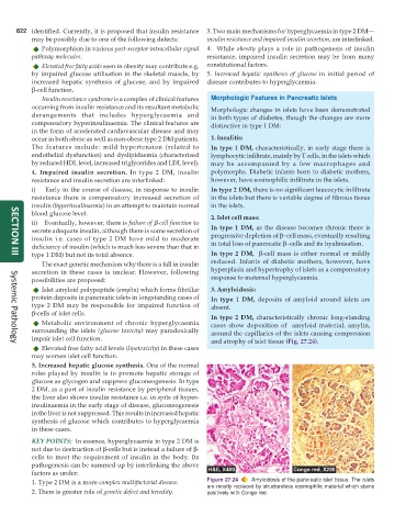
impair islet cell function. and atrophy of islet tissue (Fig. 27.24).
Systemic Pathology
Elevated free fatty acid levels (lipotoxicity) in these cases
may worsen islet cell function.
5. Increased hepatic glucose synthesis. One of the normal
roles played by insulin is to promote hepatic storage of
glucose as glycogen and suppress gluconeogenesis. In type
2 DM, as a part of insulin resistance by peripheral tissues,
the liver also shows insulin resistance i.e. in spite of hyper-
insulinaemia in the early stage of disease, gluconeogenesis
in the liver is not suppressed. This results in increased hepatic
synthesis of glucose which contributes to hyperglycaemia
in these cases.
KEY POINTS: In essence, hyperglycaemia in type 2 DM is
not due to destruction of β-cells but is instead a failure of β-
cells to meet the requirement of insulin in the body. Its
pathogenesis can be summed up by interlinking the above
factors as under:
1. Type 2 DM is a more complex multifactorial disease. Figure 27.24 Amyloidosis of the pancreatic islet tissue. The islets
are mostly replaced by structureless eosinophilic material which stains
2. There is greater role of genetic defect and heredity. positively with Congo red.