
Page 864 - Textbook of Pathology, 6th Edition
P. 864
848
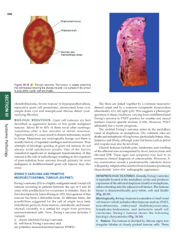
Figure 28.19 Ewing’s sarcoma. The tumour is largely extending
into soft tissues including the skeletal muscle. Cut surface of the tumour
is grey-white, cystic, soft and friable.
chondroblastoma, brown tumour of hyperparathyroidism, The three are linked together by a common neuroecto-
reparative giant cell granuloma, aneurysmal bone cyst, dermal origin and by a common cytogenetic translocation
simple bone cyst and metaphyseal fibrous defect (non- abnormality t(11; 22) (q24; q12). This suggests a phenotypic
ossifying fibroma). spectrum in these conditions varying from undifferentiated
Ewing’s sarcoma to PNET positive for rosettes and neural
SECTION III
BIOLOGIC BEHAVIOUR. Giant cell tumours are best markers (neuron-specific enolase, S-100). However, PNET
described as aggressive lesions or low grade malignant ultimately has a worse prognosis.
tumour. About 40 to 60% of them recur after curettage, The skeletal Ewing’s sarcoma arises in the medullary
sometimes after a few decades of initial resection. canal of diaphysis or metaphysis. The common sites are
Approximately 4% cases result in distant metastases, mainly shafts and metaphysis of long bones, particularly femur, tibia,
to lungs. Metastases are histologically benign and there is humerus and fibula, although some flat bones such as pelvis
usually history of repeated curettages and recurrences. Thus and scapula may also be involved.
attempts at histologic grading of giant cell tumour do not Clinical features include pain, tenderness and swelling
always yield satisfactory results. One of the factors of the affected area accompanied by fever, leucocytosis and
considered significant in malignant transformation of this elevated ESR. These signs and symptoms may lead to an
tumour is the role of radiotherapy resulting in development erroneous clinical diagnosis of osteomyelitis. However, X-
Systemic Pathology
of post-radiation bone sarcoma though primary (de novo) ray examination reveals a predominantly osteolytic lesion
malignant or dedifferentiated giant cell tumour may also with patchy subperiosteal reactive bone formation producing
occur.
characteristic ‘onion-skin’ radiographic appearance.
EWING’S SARCOMA AND PRIMITIVE MORPHOLOGIC FEATURES. Grossly, Ewing’s sarcoma
NEUROECTODERMAL TUMOUR (ES/PNET)
is typically located in the medullary cavity and produces
Ewing’s sarcoma (ES) is a highly malignant small round cell expansion of the affected diaphysis (shaft) or metaphysis,
tumour occurring in patients between the age of 5 and 20 often extending into the adjacent soft tissues. The tumour
years with predilection for occurrence in females. Since its tissue is characteristically grey-white, soft and friable
first description by James Ewing in 1921, histogenesis of this (Fig. 28.19).
tumour has been a debatable issue. At different times, the Histologically, Ewing’s tumour is a member of small round
possibilities suggested for the cell of origin have been cell tumours which includes other tumours such as: PNET,
endothelial, pericytic, bone marrow, osteoblastic, and mesen- neuroblastoma, embryonal rhabdomyosarcoma,
chymal; currently it is settled for origin from primitive lymphoma-leukaemias, and metastatic small cell
neuroectodermal cells. Now, Ewing’s sarcoma includes 3 carcinoma. Ewing’s tumour shows the following
variants: histologic characteristics (Fig. 28.20):
i) classic (skeletal) Ewing’s sarcoma; 1. Pattern. The tumour is divided by fibrous septa into
ii) soft tissue Ewing’s sarcoma; and irregular lobules of closely-packed tumour cells. These
iii) primitive neuroectodermal tumour (PNET).