
Page 865 - Textbook of Pathology, 6th Edition
P. 865
849
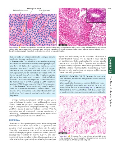
Figure 28.20 Ewing’s sarcoma. Characteristic microscopic features are irregular lobules of uniform small tumour cells with indistinct cytoplasmic
outlines which are separated by fibrous tissue septa having rich vascularity. Areas of necrosis and inflammatory infiltrate are also included. Inbox in
the right photomicrograph shows PAS positive tumour cells in perivascular location.
tumour cells are characteristically arranged around region, and infrequently in the vertebrae. Chordoma is
capillaries forming pseudorosettes. usually found in patients over the age of 40 years with no
2. Tumour cells. The individual tumour cells comprising sex predilection. Radiographically, the tumour usually
the lobules are small and uniform resembling lymphocytes appears as an osteolytic lesion. Symptoms of spinal cord
and have ill-defined cytoplasmic outlines, scanty compression may be present. The tumour grows slowly and CHAPTER 28
cytoplasm and round nuclei having ‘salt and pepper’ infiltrates adjacent structures but metastases develop rarely.
chromatin and frequent mitoses. Based on these Recurrences after local excision are frequent and the tumour
cytological features the tumour is also called round cell almost invariably proves fatal.
tumour or small blue cell tumour. The cytoplasm contains MORPHOLOGIC FEATURES. Grossly, the tumour is
glycogen that stains with periodic acid-Schiff (PAS) soft, lobulated, translucent and gelatinous with areas of
reaction. The consistently expressed cell surface marker haemorrhages.
by tumour cells of ES/PNET group is CD99 which is a Microscopically, chordoma is composed of highly vacuo-
product of MIC-2 gene located on X and Y chromosome. lated physaliphorous cells surrounded by a sea of
3. Other features. The tumour is richly vascularised and intercellular mucoid material (Fig. 28.21). Histologic
lacks the intercellular network of reticulin fibres. There differentiation between chordoma and chondrosarcoma
may be areas of necrosis and acute inflammatory cell or mucin-secreting carcinoma may sometimes be difficult. The Musculoskeletal System
infiltration. Focal areas of reactive bone formation may
be present.
Ewing’s sarcoma metastasises early by haematogenous
route to the lungs, liver, other bones and brain. Involvement
of other bones has prompted a suggestion of multicentric
origin of Ewing’s sarcoma. The prognosis of Ewing’s sarcoma
used to be dismal (5-year survival rate less than 10%). But
currently, use of combined regimen consisting of
radiotherapy and systemic chemotherapy has improved the
outcome greatly (5-year survival rate 40-80%).
CHORDOMA
Chordoma is a slow-growing malignant tumour arising from
remnants of notochord. Notochord is the primitive axial
skeleton which subsequently develops into the spine.
Normally, remnants of notochord are represented by
notochordal or physaliphorous (physalis = bubble, phorous =
bearing) cells present in the nucleus pulposus and a few
clumps within the vertebral bodies. Chordomas thus occur Figure 28.21 Chordoma. The tumour cells are quite variable in size
having characteristic bubbly cytoplasm (physaliphorous cells) and
in the axial skeleton, particularly sacral and spheno-occipital anisonucleocytosis. The background is myxoid.